
Inner Foot Anatomy: What You Need to Know About the Body's Most Complex Structure
The anatomy of the inner foot: 26 bones, 33 joints and more than 100 muscles, ligaments and tendons. How they work together and why problems arise.
By Dr. Luigi ManziUpdated on
Have you ever wondered why your foot hurts so much when it gets inflamed? Or why certain areas seem more tender than others?
The answer lies in the anatomy of the inner foot. An incredibly complex structure that we often underestimate. Consider that this small space houses 26 bones, 33 joints, and more than 100 muscles, ligaments, and tendons. It's no coincidence that many orthopedic problems arise right here.
In my office, I constantly see patients who come in with inner foot pain, inner lateral foot pain, or general discomfort in the medial aspect of the foot. And I often find they have no idea what's going on "down there." That's why I've decided to explain everything you need to know.
Bones: The Scaffolding of Your Foot
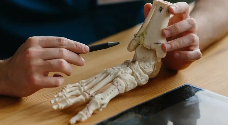
Let's start with the basics. The foot is divided into three main zones: the forefoot (with the toes), the midfoot (the central part), and the hindfoot (heel and ankle).
The tarsal bones are seven and form the basis of everything. The calcaneus is the largest—the one you stand on when walking. Then there's the talus, which connects to the ankle.
The metatarsal, on the other hand, is made up of five long bones that connect the middle of the foot to the toes. So, when you feel pain in this area of the inside of your foot, the first metatarsal—the one in the big toe—is often involved. Finally, the phalanges are the bones of the toes.
Each bone has its own specific role. The heel absorbs impact. The talus distributes weight. The metatarsals propel you forward when you walk. It's a perfect system when it works well.
Muscles: the hidden protagonists
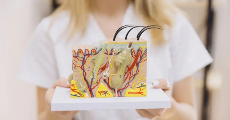
The muscles of the foot are divided into two categories: intrinsic muscles, which are entirely inside the foot, and extrinsic muscles, which originate from the leg.
The intrinsic muscles are small but crucial. They control the fine movements of the toes, maintain the arch of the foot, and stabilize everything during walking. When these muscles weaken—which often happens with age or a sedentary lifestyle—problems begin.
The extrinsic muscles, on the other hand, are the "big movers." The tibialis anterior allows you to lift your toes. The peroneals stabilize the ankle laterally. The tibialis posterior—this is very important—supports the medial arch. When the tibialis posterior isn't working properly, pain often occurs on the inside of the foot.
The Achilles tendon deserves a special mention. It's the strongest tendon in the human body, but also one of the most prone to problems. It connects the calf to the heel and gives you the power to walk, run, and jump.
Joints: Where Everything Moves
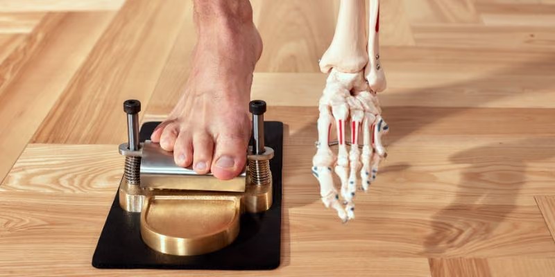
The foot has numerous joints. The most important is undoubtedly the ankle joint—the one that connects to the ankle itself. It primarily allows for flexion and extension of the foot.
But the most delicate joints are those of the midfoot. The talonavicular joint is crucial for the longitudinal arch. When it "breaks down," the entire foot collapses inward.
The metatarsophalangeal joints—the joints between the long bones of the foot and the toes—are subjected to enormous stress during walking. It's no coincidence that problems like hallux valgus and hallux rigidus arise here.
Ligaments and tendons: the support network
The ligaments of the foot form an incredibly dense network. The deltoid ligament, on the inside of the ankle, is one of the strongest. When it tears, the ankle becomes unstable.
The plantar fascia is probably the most famous structure—in the sense that when it hurts, everyone notices. It's the fibrous band that runs from the heel to the toes and supports the arch of the foot. When it becomes inflamed, plantar fasciitis occurs.
The tendons of the intrinsic muscles create a dynamic support network. The posterior tibialis tendon, in particular, is crucial for maintaining the medial arch. Its dysfunction is one of the main causes of chronic lateral instep pain.
Nerves and vessels: the information highway

Three main nerves pass through the foot: the tibial, deep peroneal, and superficial peroneal nerves. The tibial nerve passes behind the medial malleolus and innervates most of the intrinsic muscles.
When this nerve becomes compressed in the tarsal tunnel—a sort of "carpal tunnel" of the foot—pain, tingling, and numbness appear on the inside of the foot. It's a problem I often see in athletes.
The vascular system of the foot is delicate. The posterior tibial artery and the dorsalis pedis artery carry blood, while venous return depends on the superficial and deep veins. Circulatory problems can cause swelling, night cramps, and changes in sensation.
The main functions of the foot

The foot isn't just a static support. During walking, it becomes a complex lever that must absorb the impact of the heel, adapt to the terrain, support the entire body weight, and propel the body forward.
This continuous transformation requires perfect coordination between all structures. When even one of these changes, the entire system is affected.
Most common pathologies
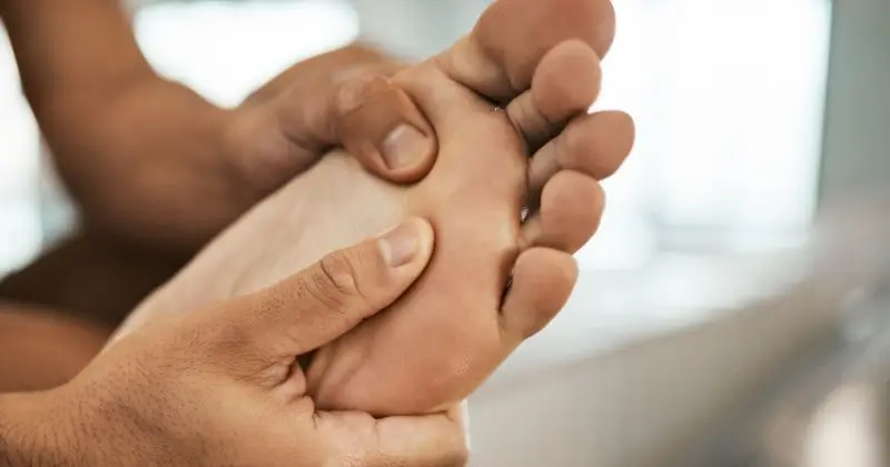
In my office I mainly see: hallux valgus (that “onion” that sticks out), hallux rigidus (when the joint locks up), plantar fasciitis (pain under the heel), Achilles tendinitis, metatarsalgia (pain under the toes).
Each pathology has its own specific causes, but they often stem from overloading, improper footwear, and postural alterations. Flat feet and high arches, for example, completely alter weight distribution.
Prevention: What you can do
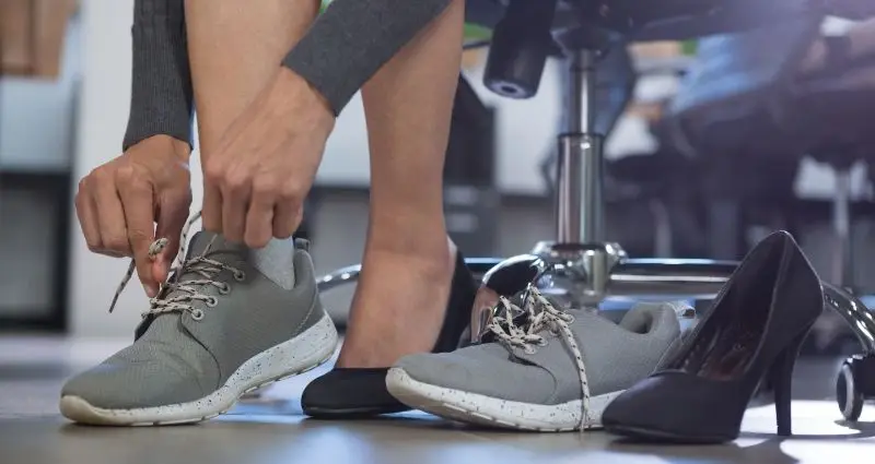
The right shoes make all the difference. They must support the arch, provide cushioning, and have the correct size. Stretching exercises for the calf and plantar fascia are essential. Weight control reduces overload.
Regular physical activity keeps all the muscles in your feet active. Even simply walking barefoot around the house helps keep your intrinsic muscles strong.
SmartHallux: Where we can help you

Here at SmartHallux, we specialize in foot and ankle surgery. Dr. Luigi Manzi, who has extensive experience in this field, provides each patient with a personalized treatment plan using the most modern techniques.
The SmartHallux team treats conditions such as hallux valgus , Morton's neuroma , and other deformities that cause foot pain. Providing comprehensive support throughout the treatment process, SmartHallux can be a winning choice for effective treatment tailored to your needs. Book your specialist consultation with the SmartHallux team now (opens in a new tab).
Questions you often ask me
Why do I feel pain inside my foot in the morning?
It's often related to the plantar fascia, which "shortens" during the night and stretches sharply when you take your first step. But there may be other causes that need to be evaluated.
Is foot surgery always necessary?
Absolutely not. Many problems are resolved with conservative therapies. I consider surgery when the pain significantly limits quality of life and conservative treatments have not worked.
How long does recovery take after foot surgery?
It depends on the type of surgery. For a simple bunion, it takes 4-6 weeks to return to normal walking. For more complex surgeries, the timeframe may be longer, but each case is different.